QCM-I is a powerful technique for measuring the adsorption or binding of polymers, proteins, nano-particles and other molecules to a surface. Multi-parametric data is obtained in real time, detailing changes of the hydrated mass and rigidity of layers coupled to the sensor surface. Here, the mass and viscoelastic changes can be understood for a range of adsorptive events, including the polymer modification of a surface, followed by the physisorption of biotin-BSA, binding of streptavidin functionalized quantum dots and further in-filling with streptavidin.
Experimental Introduction
The experiment was performed using a MicroVacuum ITO coated QCM-I sensor crystal, with a continuous flow of HEPES buffer, pH7.4, over the sensor surface, Samples were introduced from a fixed volume injection loop, followed by rinsing with the running HEPES buffer. Consecutive sample solutions injected were: 2M NaCl (to provide a purely viscous signal change), polyethylenimine (PEI) solution (polymer adsorption), PBS (to compress the PEI layer), biotin-BSA (protein physisorption), 3x Streptavidin-Quantum Dot(20nm nano-particle from Invitrogen, specific binding to biotin) and streptavidin (to in-fill around the SA-QD).
The QCM-I instrument monitors the impedance spectrum of the oscillating quartz crystal sensor, tracking the changes in the resonant frequencies of its fundamental and overtones as well as their peak widths (FWHM or w, all in Hz). The former is a function of the hydrated mass coupled to the sensor surface and the latter, of the viscoelasticity or rigidity of the coupled material.
Results
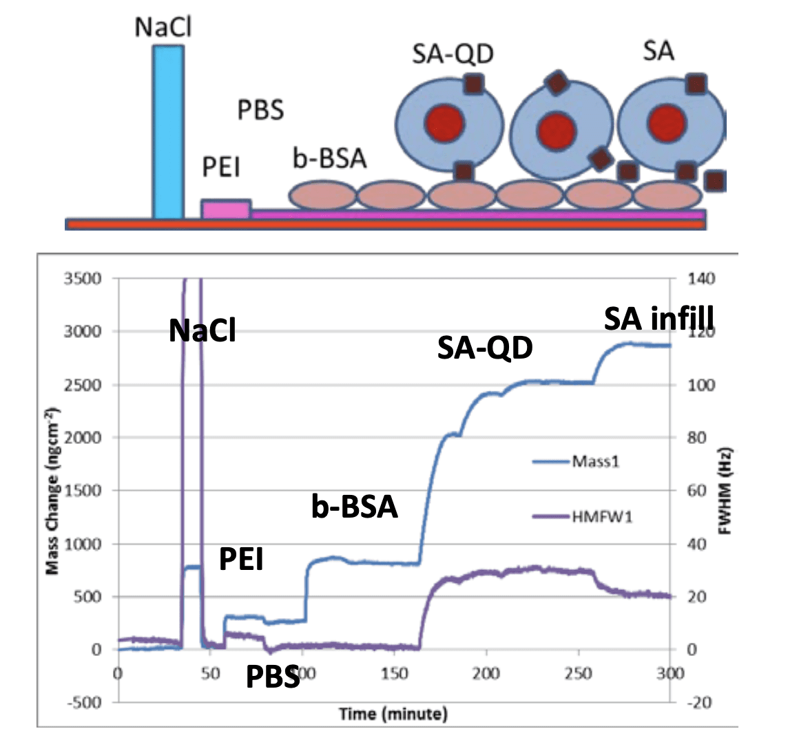
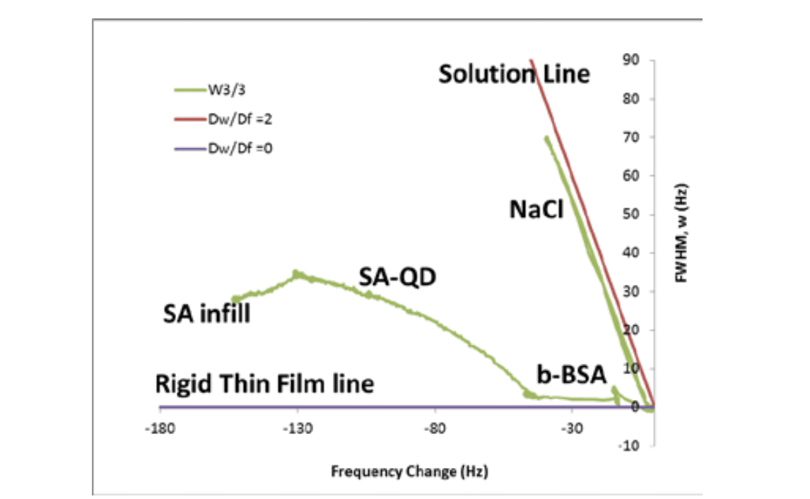
Figure 1 shows a schematic cartoon of the experiment and the data obtained. The fundamental frequency change has been converted to mass using the Sauerbrey equation1 which assumes a rigid coupled layer, which includes the mass of water trapped within the film. If the film is rigid, the peak width is unchanged. For a very viscous layer, a large width change will be observed. This can be seen for the 2M NaCl injection, where a large transient width change is observed for a small “mass” change, as this is solely a bulk solution exchange and not a surface process. This can also be seen in a plot of Width change vs. Frequency change in Figure 2. For a purely viscous solution, ∆w/∆f=2. For a purely rigid layer ∆f/∆w=0.
PEI: Injection of PEI adsorbs a ~300ngcm-2 viscoelastic hydrated polymer layer to the surface. This becomes rigid and expels some water as PBS buffer is injected, due to the multivalent phosphate ions binding into the amino-polymer.
b-BSA: The biotinylated protein adsorbs electrostatically as a rigid film onto the PEI. Assuming a density of 1g/ml, the ~550ngcm-2 hydrated layer has a thickness of 5.5nm.
SA-QD: The initial SA-QD binding causes a relatively rapid FWHM increase as the ~20nm particle is attached via a single SA anchor point and so is not rigid. As binding progresses and the surface fills in, the whole layer becomes more rigid and the mass increases with less width change.
SA: Free streptavidin which binds to any still available b-BSA, fills the gaps in-between the SA-QD. This reduces the layer flexibility and causes the coupled mass to increase and the FWHM to decrease, closer to the rigid film line in figure 2. The combined SA-QD and SA mass change of 2100ngcm-2 is indeed close to that expected for a layer of ~20nm nano- particles surrounded by water.
Reference: 1 Using Complementary Acoustic and Optical Techniques for Quantitative Monitoring of Biomolecular Adsorption at Interfaces, R.Konradi, M.Textor and E.Reimhult, Biosensors 2012, 2, 341-376
Experiments performed by Dr M Swann in the lab of Prof. J.Lu, University of Manchester, UK.